
The Muscular System: Exploring the Magnificent Muscles of the Human Body
How many muscles are there in the human body and how are they arranged? This guide is designed to be a useful resource, providing you with the names of each muscle, categorized groupings, and accompanying images for visual reference. Whether you're a student, a health professional, or simply someone with a curiosity about human anatomy, this post will serve as a valuable reference, enabling you to expand your knowledge and explore the intricate details of each muscle.
Anterior

Posterior

There are approximately 650 to 700 muscles in the human body, although the exact number depends on how ‘muscle’ is defined. All the muscles may be arranged into four major groups, with sub-divisions and further sub-divisions:
1. Head Zone
Head
-
Orbitalis
Superior rectus
Inferior rectus
Medial rectus
Lateral rectus
Common tendinous ring
Superior oblique
Inferior oblique
Levator palpebrae superioris
-
Tensor tympani
Stapedius
Mucosa of tympanic cavity
-
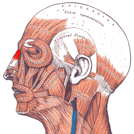
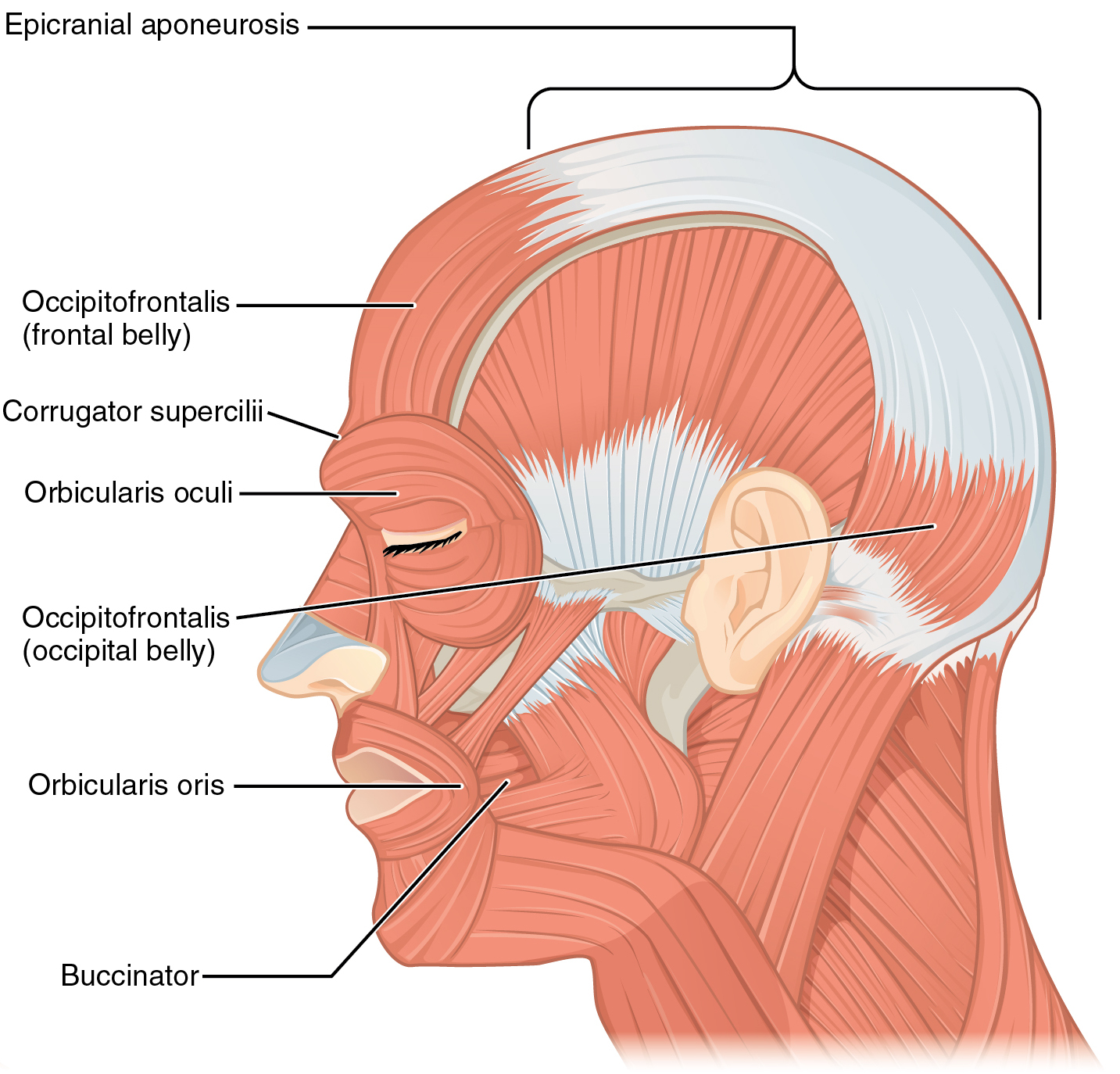
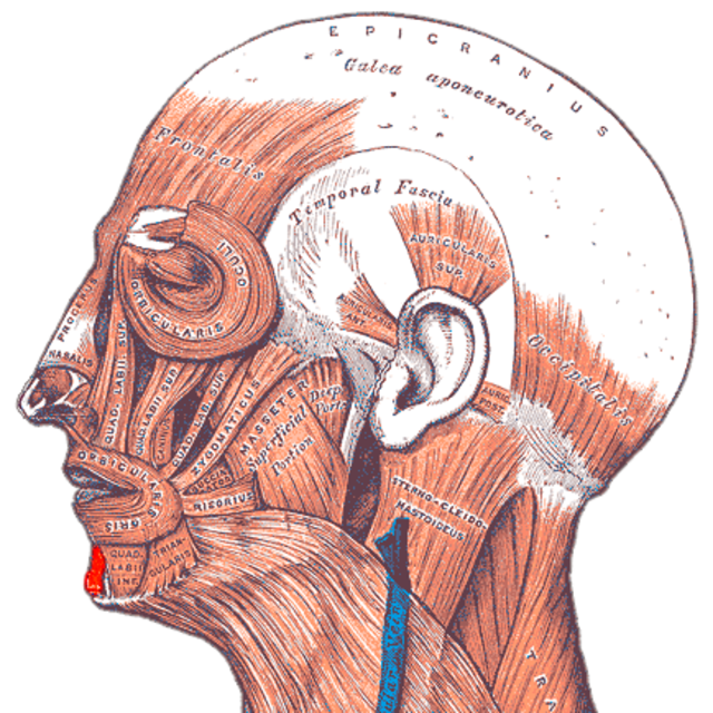
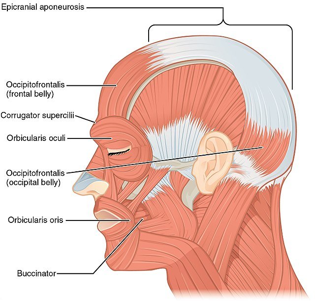
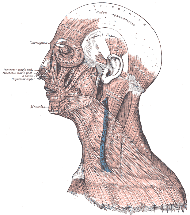
Epicranius
Procerus
Nasalis
Depressor septi nasi
Orbicularis oculi
Corrugator supercilii
Depressor supercilii
Auricularis anterior
Auricularis superior
Auricularis posterior
Orbicularis oris
Depressor anguli
Transversus menti
Risorius
Zygomaticus major
Zygomaticus minor
Levator labii superioris
Levator labii superioris alaeque nasi
Depressor labii inferioris
Levator anguli oris
Modiolus
Buccinator
Mentalis
-
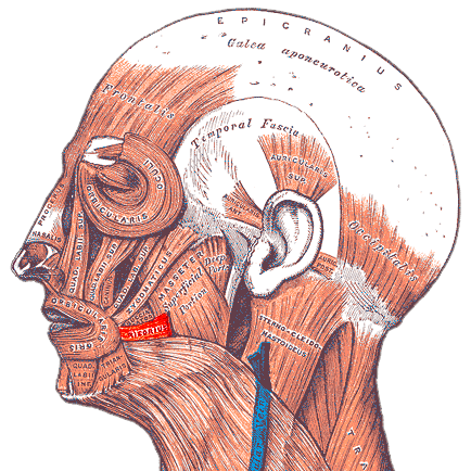
Masseter
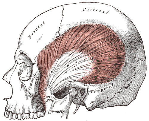
Temporalis
Lateral pterygoid
Medial pterygoid
Buccopharyngeal fascia
Masseteric fascia
Parotid fascia
Temporal fascia
-
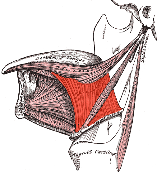
Genioglossus
Hyoglossus
Styloglossus
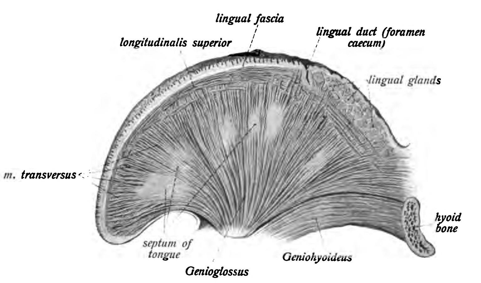
Superior longitudinal muscle
Inferior longitudinal muscle
Transverse muscle
Vertical muscle
Palatoglossus
-
Palatine aponeurosis
Levator veli palatini
Tensor veli palatini
Musculus uvulae
Palatoglossus
Palatopharyngeus


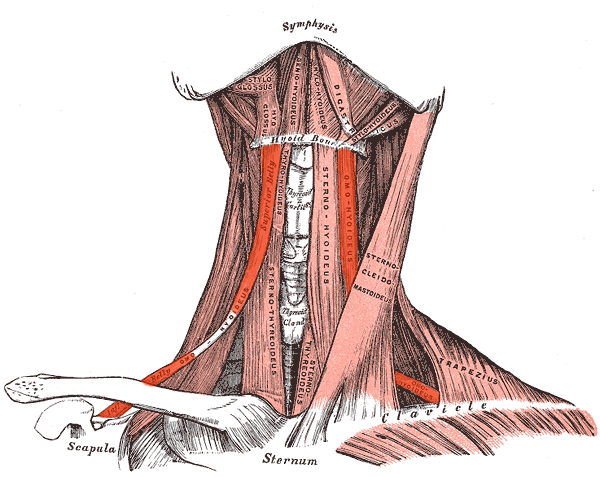
Neck
-
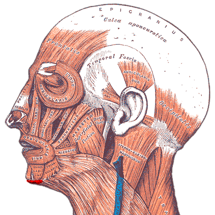
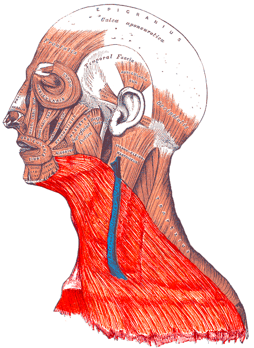
Platysma
Longus colli
Longus capitis
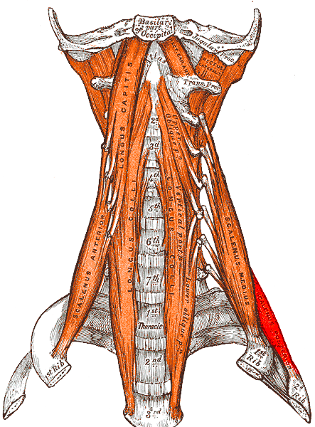
Scalenus anterior
Scalenus medius
Scalenus posterior
(Scalenus minimus)
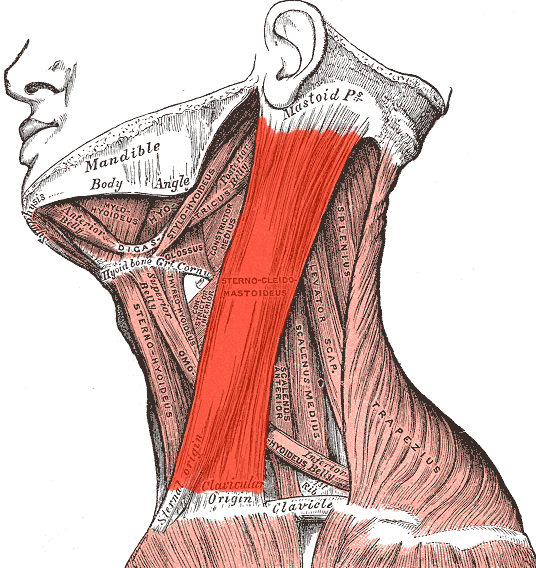
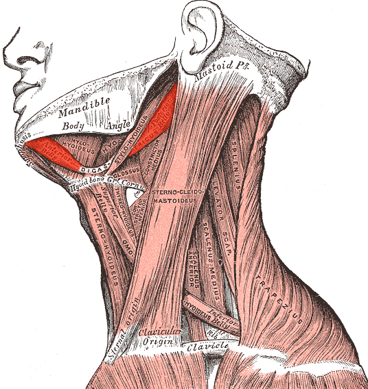
Sternocleidomastoid
-
Rectus capitis anterior
Rectus capitis lateralis
Rectus capitis posterior major
Rectus capitis posterior minor
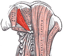
Obliquus capitis superior
Obliquus capitis inferior
-
Digastric
Stylohyoid
Mylohyoid
Geniohyoid
-
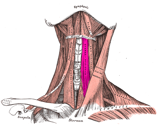
Sternohyoid
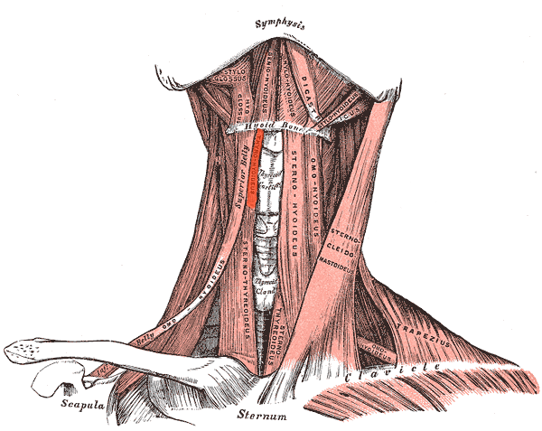
Omohyoid
Sternothyroid
Thyrohyoid
(Levator glandulae thyroideae)
-
Investing layer
Pretracheal layer
Prevertebral layer
Carotid sheath
-
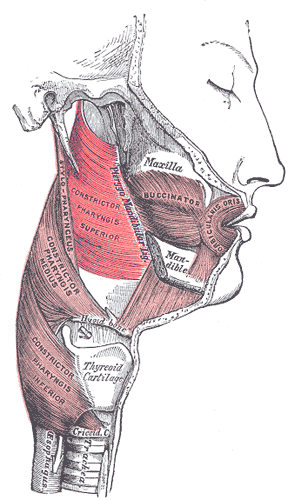
Pharyngeal raphe
Pterygomandibular raphe
Superior constrictor
Middle constrictor
Inferior constrictor
Stylopharyngeus
Salpingtopharyngeus
Palatopharyngeus
Buccopharyngeal fascia
Peripharyngeal space
-
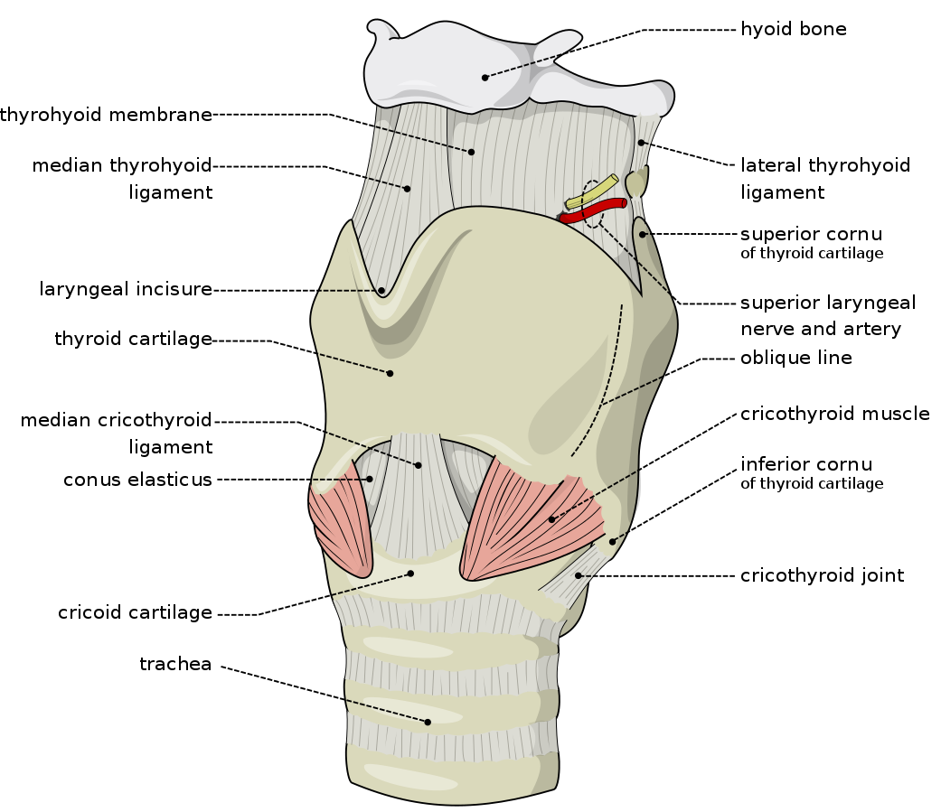
Cricothyroid
Posterior crico-arytenoid
(ceratocricoid)
Lateral crico-arytenoid
Vocalis
Thyro-arytenoid
Oblique arytenoid
Transverse arytenoid


2. Back Zone
-
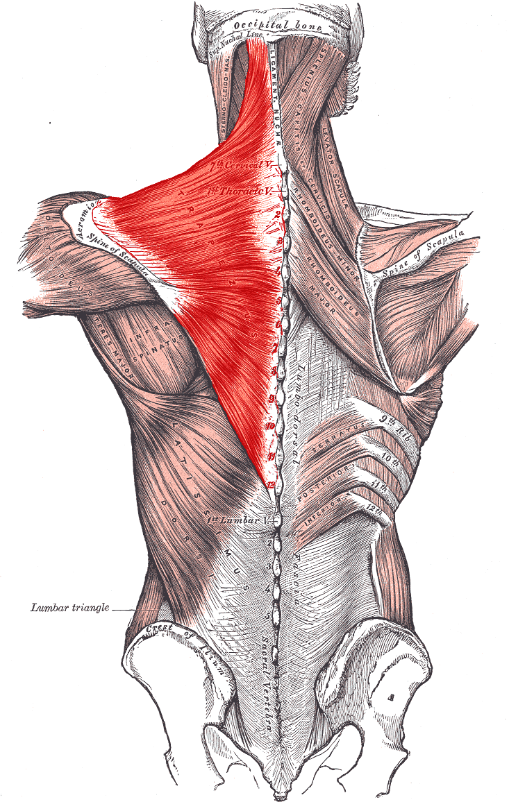
Trapezius
(Transversus nuchae)
Latissimus dorsi
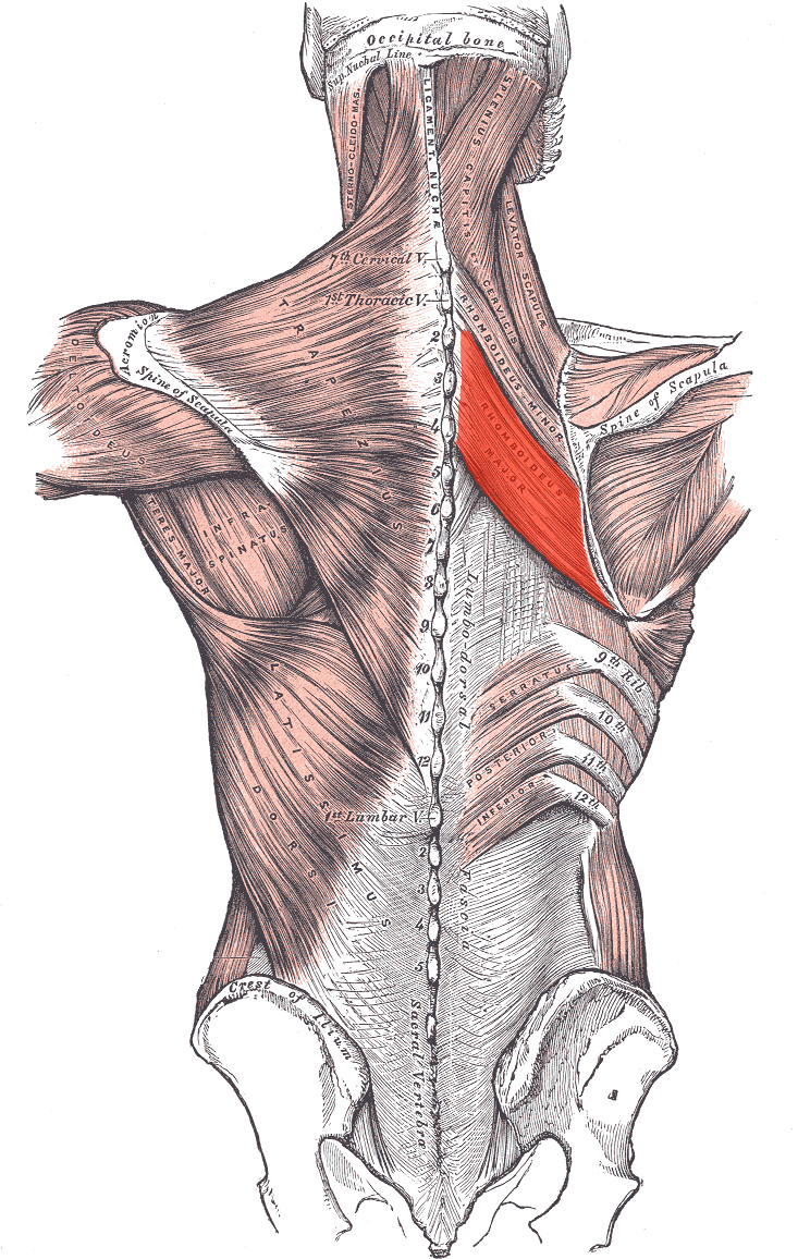
Rhomboid major
Rhomboid minor
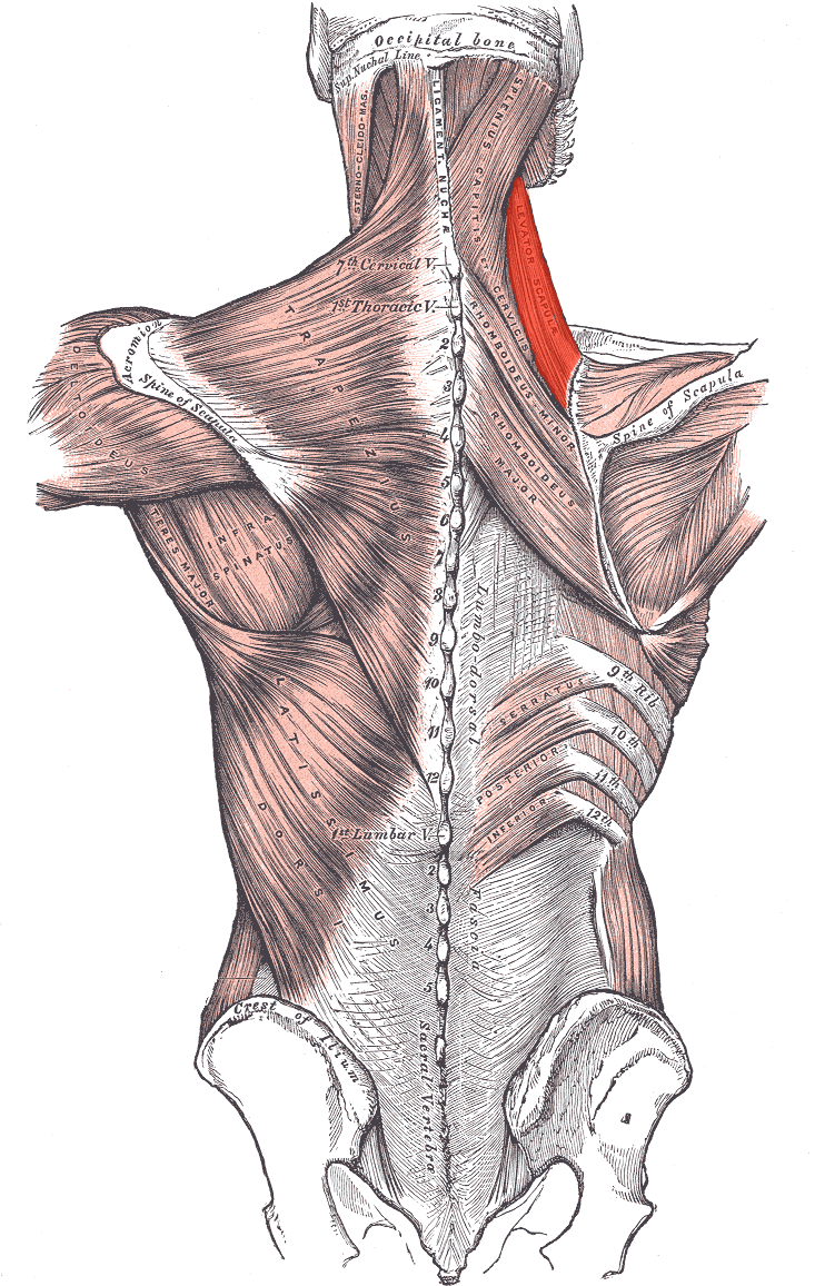
Levator scapulae
Serratus posterior inferior
Serratus posterior superior
Anterior cervical intertransversarii
Lateral posterior cervical intertransversarii
Intertransversarii laterals lumborum
Nuchael fascia
-
Erector Spinae
Spino-transversales
Transverso-spinales
Inter-spinales
Inter-transversarii
Throacolumbar fascia


3. Front Zone
-
(Sternals)
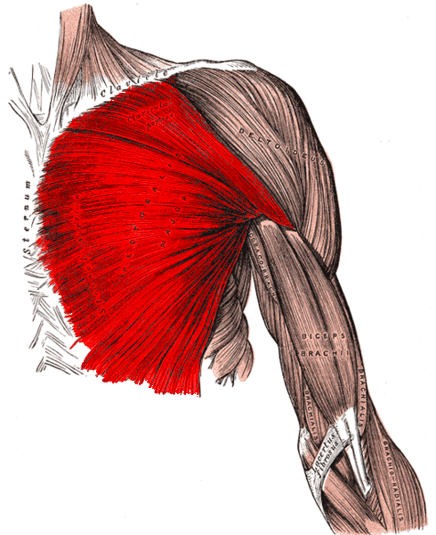
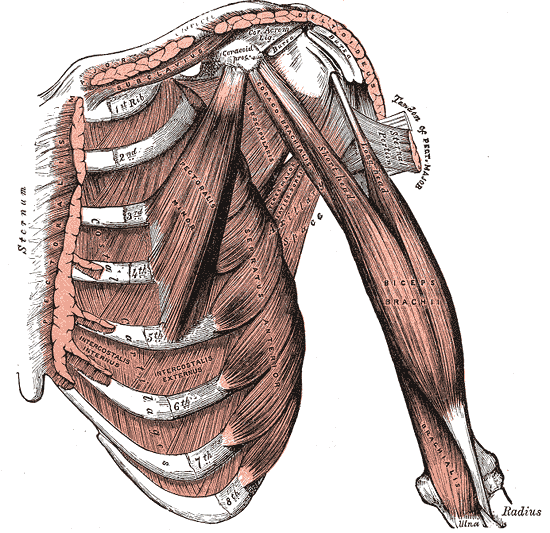
Pectoralis major
Pectoralis minor
Subclavius
Serratus anterior
Levatores costarum
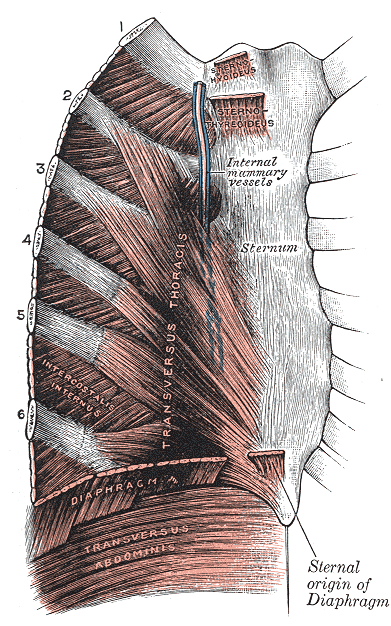
External intercostal muscle
Internal intercostal membrane
Internal intercostal muscle
Internal intercostal membrane
Innermost intercostal muscle
Subcostales
Transversus throacis
Pectoral fascia
Clavipectoral fascia
Thoracic fascia
Endothoracic fascia
Diaphragm
-
Abdomen
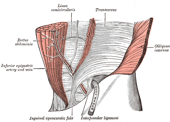
Rectus abdominis
Pyramidalis
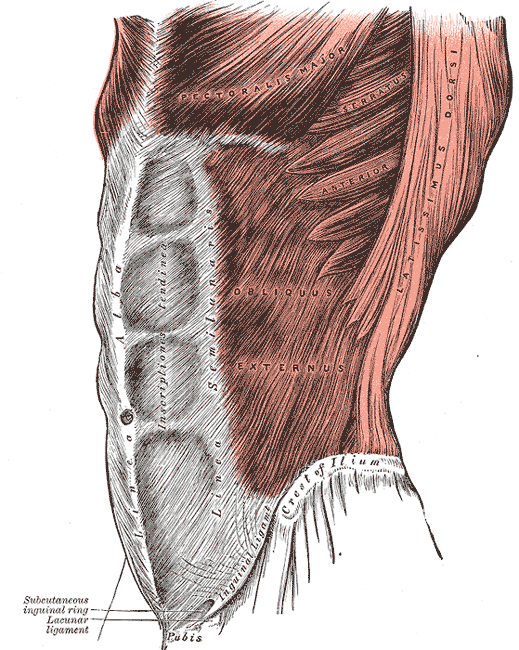
External oblique
Superficial inguinal ring
Internal oblique
Transversus abdominis
Linea alba
Linea semilunaris
Inguinal canal
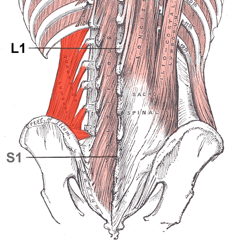
Quadratus lumborum
Abdominal fascia
Pelvic fascia
Pelvic diaphragm
Perineal


4. Limbs
-
Deltoid
Supraspinatus
Infraspinatus
Teres minor
Teres major
Subscapularis
Biceps brachii
Coracobrachialis
Brachialis
Triceps brachii
Anconeus
Articulasis cubiti
Pronator teres
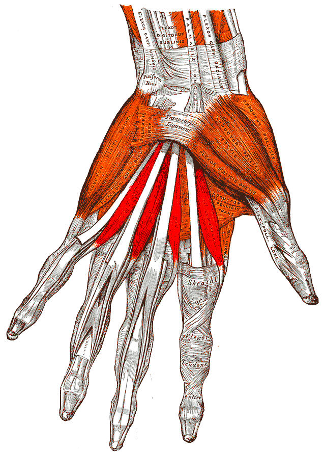
Flexor carpi radialis
Palmaris longus
Flexor carpi ulnaris
Flexor digitorum profundus
Flexor pollicis longus
Pronator quardratus
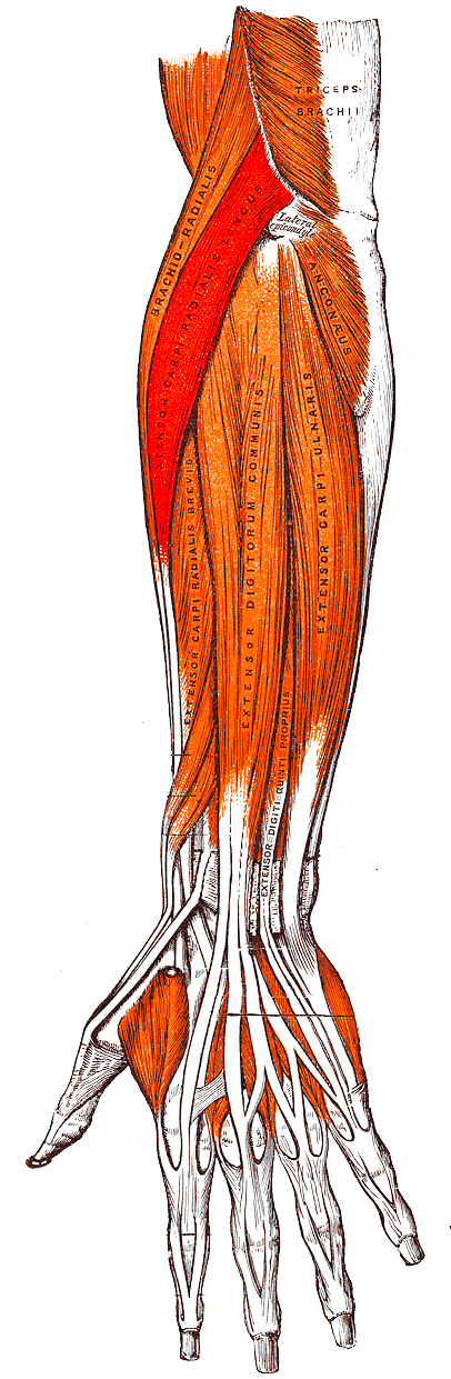
Brachioradialis
Extensor carpi radialis longus
Extensor carpi radialis brevis
Extensor digitorum
Extensor digiti minimi
Extensor carpi ulnaris
Supinator
Abductor pollicis longus
Extensor pollicis brevis
Extensor pollicis longus
Extensor indicis
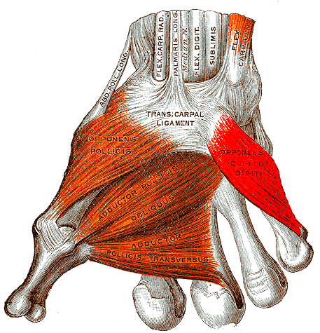
Palmaris brevis
Abductor pollicis brevis
Flexor pollicis brevis
Opponnens pollicis
Adductor pollicis
Abductor digiti minimi
Flexor digiti minimi brevis
Opponens digiti minimi
Lumbricales
Dorsal interossei
Palmar interossei
-
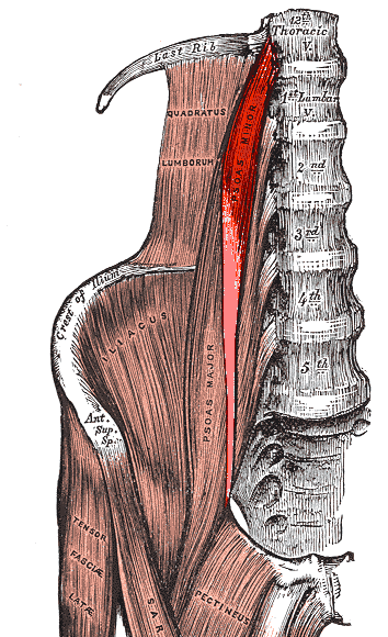
Iliopsoas
(Psoas minor)
Gluteus maximus
Gluteus medius
Gluteus minimus
Gluteal aponeurosis
Tensor fascia latae
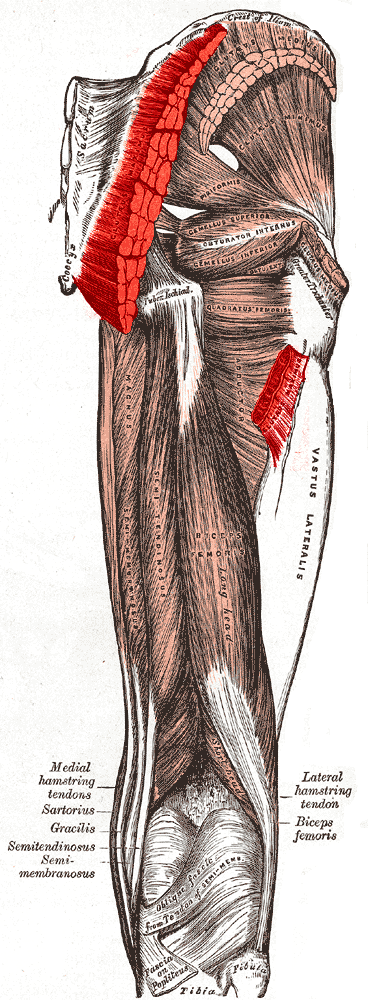
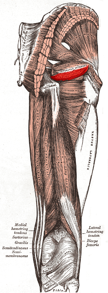
Piriformis
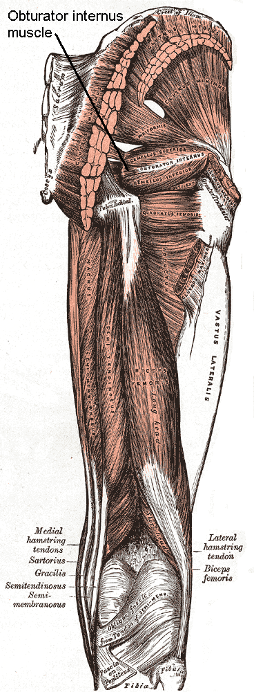
Obturator internus
Gemellus superior
Gemellus inferior
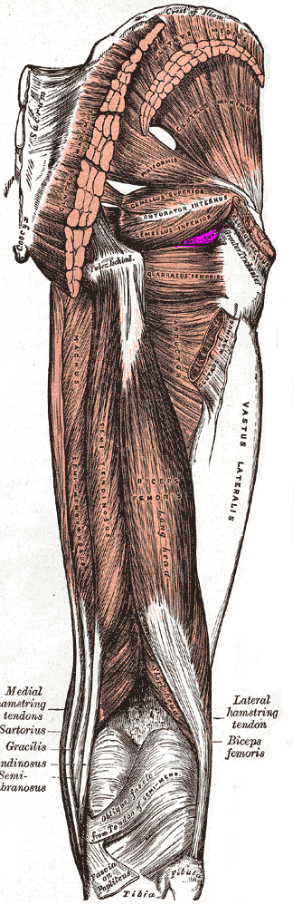
Quadratus femoris
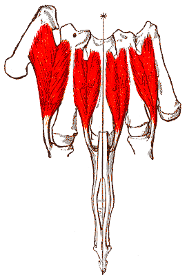
Sartorius
Quadriceps femoris
Articuloris genus
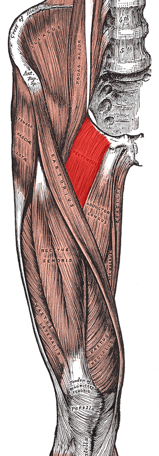
Pectineus
Adductor longus
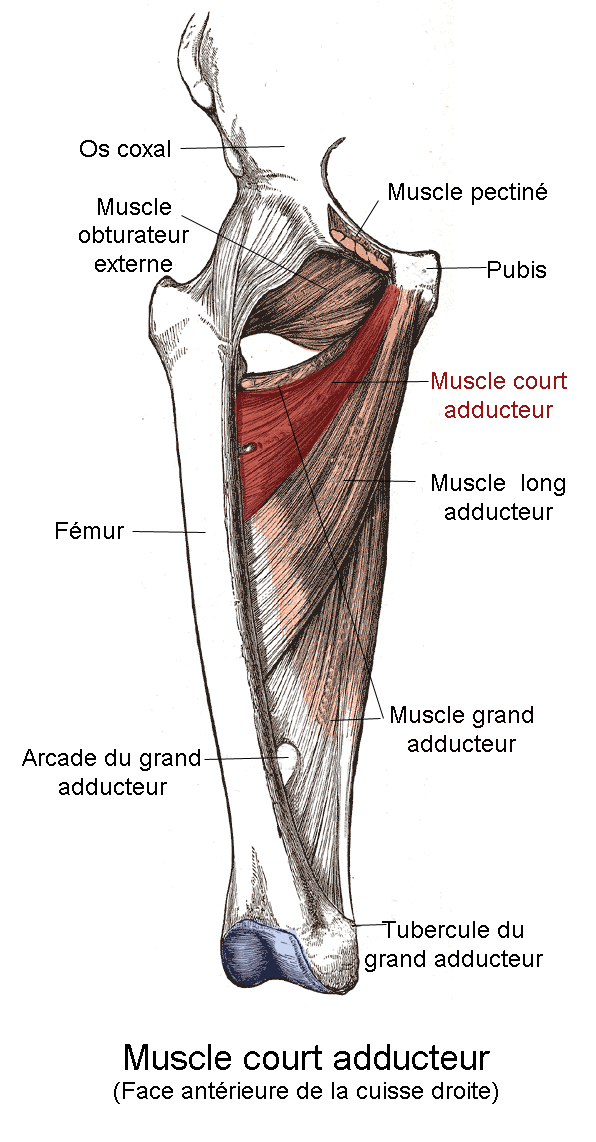
Adductor brevis
Adductor magnus
Adductor minimus
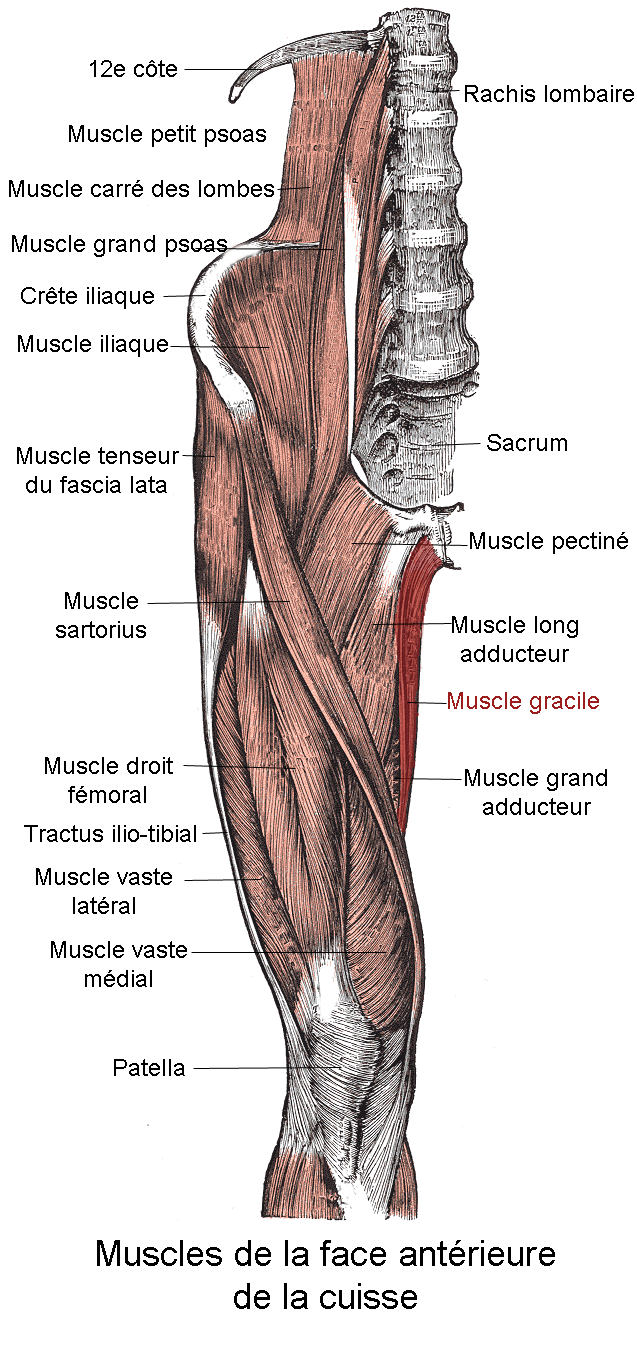
Gracilis
Obturator externus
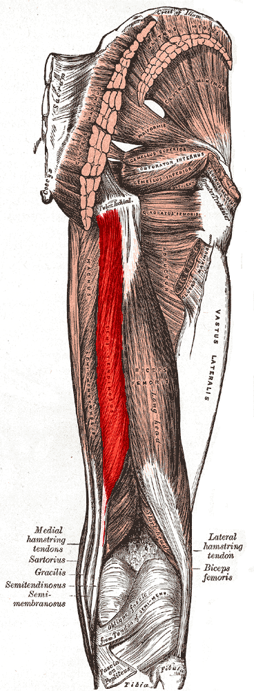
Biceps femoris
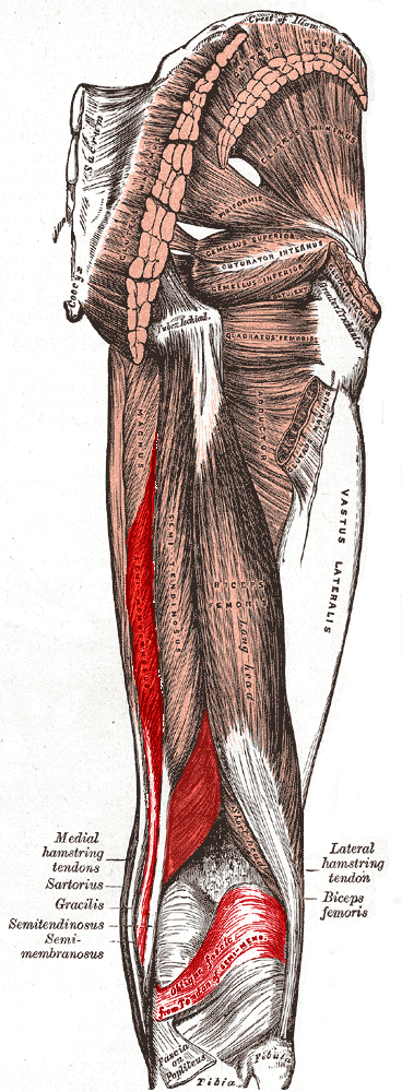
Semitendinosus
Semimembranosus
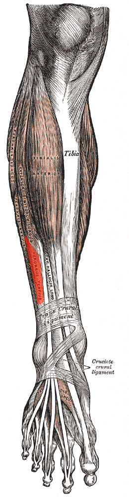
Tibialis anterior
Extensor digitorum longus
Fibularis tertius
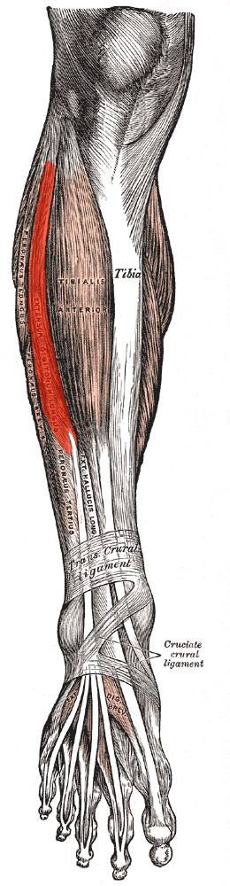
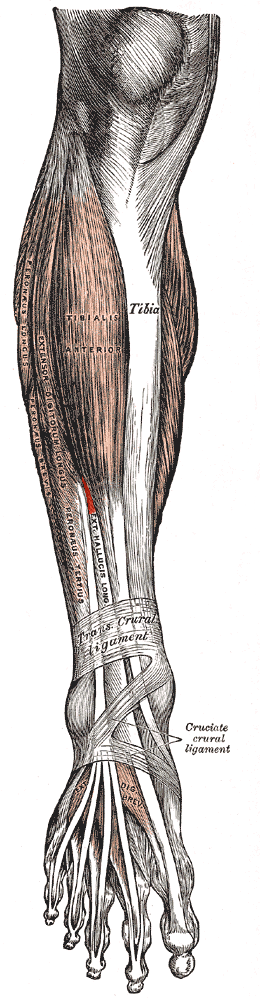
Extensor hallucis longus
Fibularis longus
Fibularis brevis
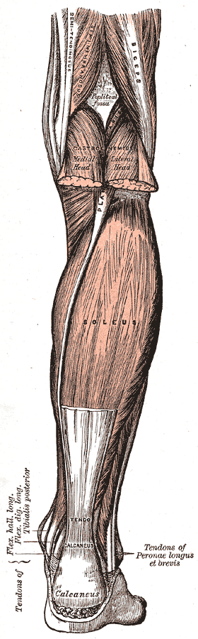
Triceps surae
Plantaris
Popliteus
Tibialis posterior
Flexor digitorum longus
Flexor hallucis longus
Extensor hallucis brevis
Extensor digitorum brevis
Abductor hallucis
Flexor hallucis brevis
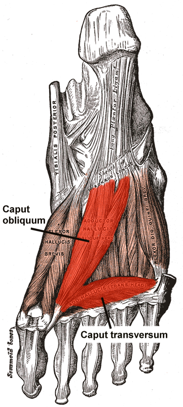
Adductor hallucis
Abductor digiti minimi
(Opponens digiti minimi)
Flexor digiti minimi brevis
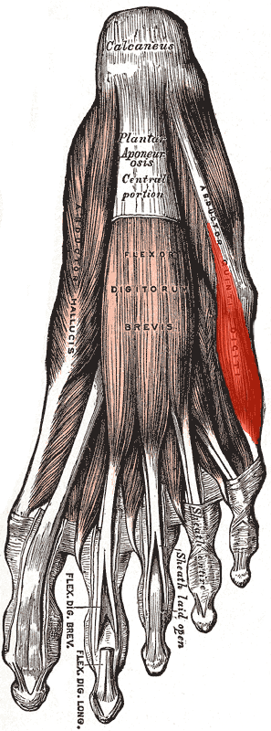
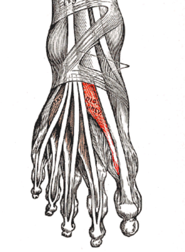
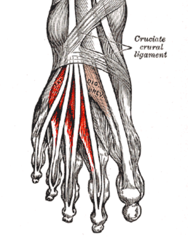
Flexor digitorum brevis
Quadratus plantae
Lumbricals
Dorsal interossei
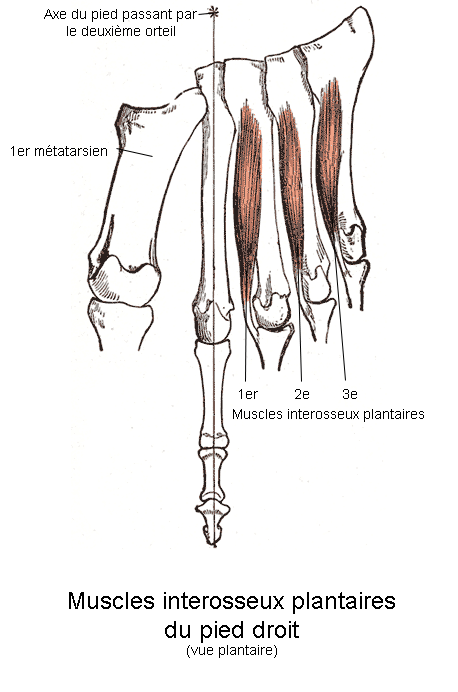
Plantar interossei


Muscle Actions
-
Head
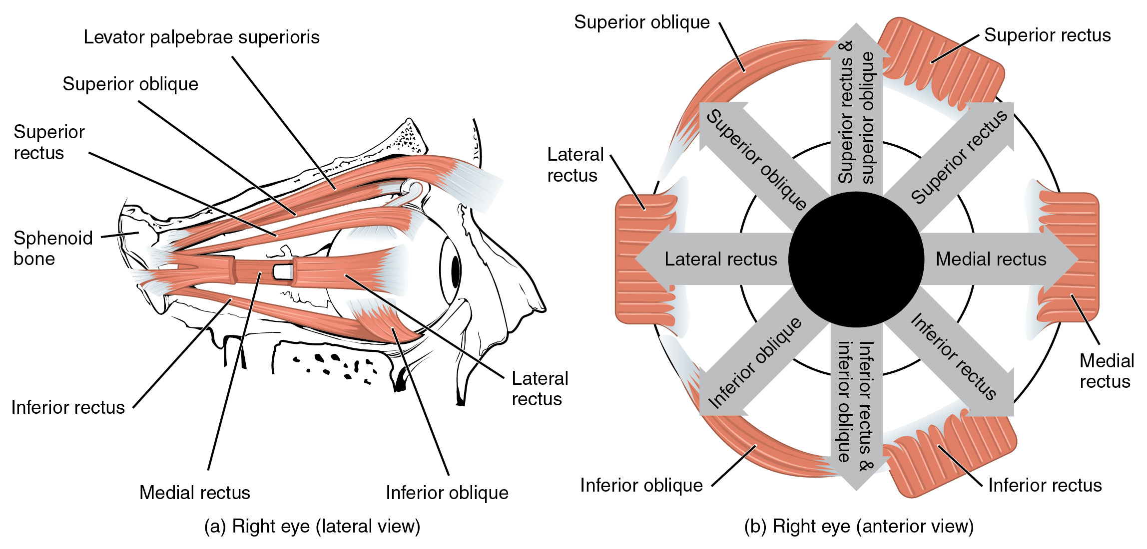
Extra-ocular (Eyes)
Orbitalis: Protrudes the eyeball (minimal functional effect in humans).
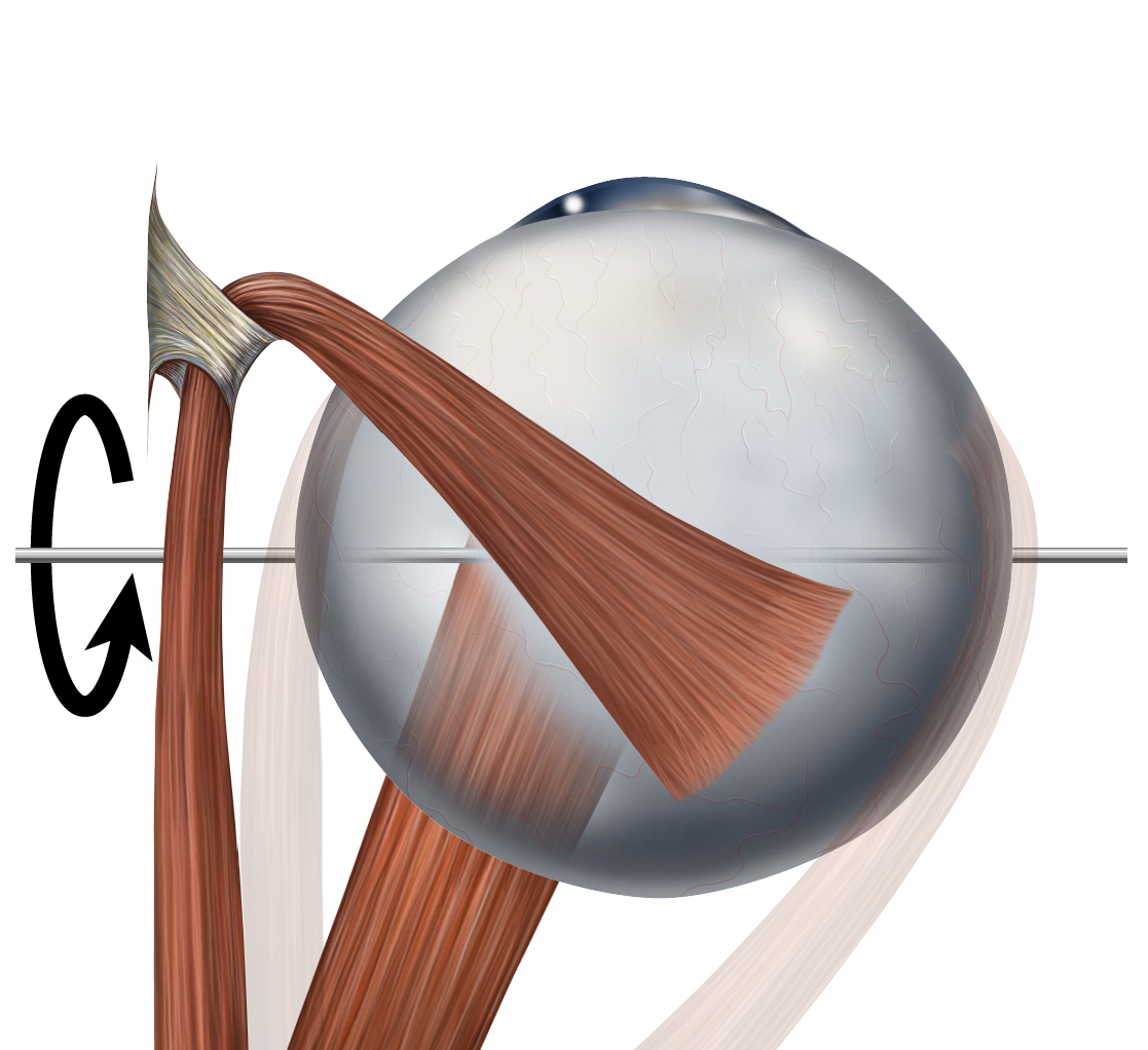
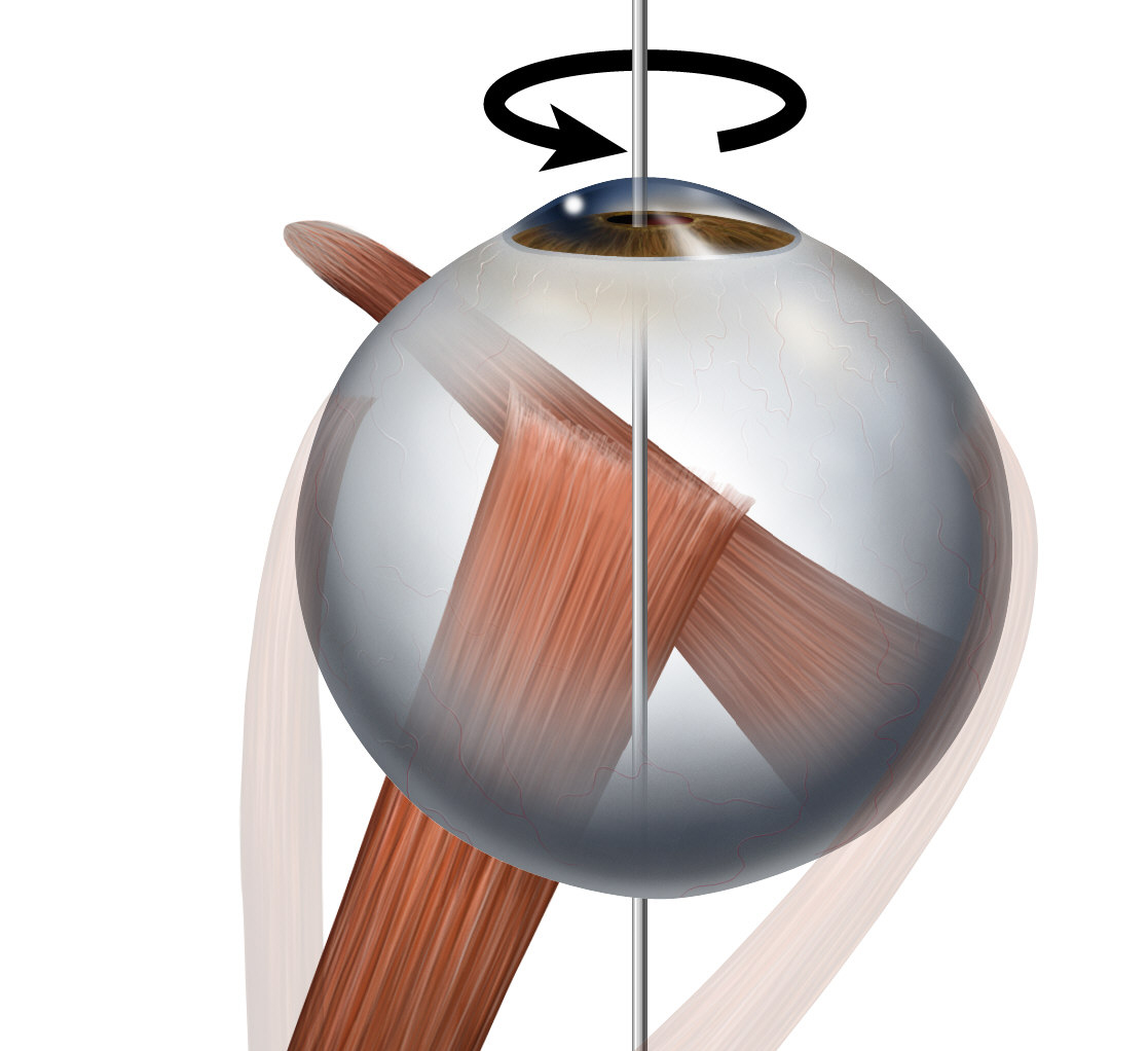
Superior rectus: Elevates, adducts, and medially rotates the eyeball.
Inferior rectus: Depresses, adducts, and laterally rotates the eyeball.
Medial rectus: Adducts the eyeball (moves it toward the nose).
Lateral rectus: Abducts the eyeball (moves it toward the ear).
Common tendinous ring: * Superior oblique: Depresses, abducts, and medially rotates the eyeball.
Inferior oblique: Elevates, abducts, and laterally rotates the eyeball.
Levator palpebrae superioris: Elevates the upper eyelid.
Auditory Ossicles (Ears)
Tensor tympani: Tenses the tympanic membrane to reduce sound transmission.
Stapedius: Stabilizes the stapes bone to dampen loud vibrations.
Mucosa of tympanic cavity: #### Facial
Epicranius (Occipitofrontalis): Raises eyebrows and wrinkles the forehead.
Procerus: Pulls down the skin between the eyebrows (frowning).
Nasalis: Flares and compresses the nostrils.
Depressor septi nasi: Depresses the nasal septum.
Orbicularis oculi: Closes the eyelids (blinking and squinting).
Corrugator supercilii: Draws eyebrows together medially (vertical frowning).
Depressor supercilii: Depresses the eyebrows.
Auricularis anterior: Moves the ear forward.
Auricularis superior: Elevates the ear.
Auricularis posterior: Moves the ear backward.
Orbicularis oris: Closes and protrudes (purses) the lips.
Depressor anguli: Pulls the corners of the mouth downward.
Transversus menti: Assists in depressing the corners of the mouth.
Risorius: Draws the corners of the mouth laterally (grimacing).
Zygomaticus major: Pulls corners of mouth upward and outward (smiling).
Zygomaticus minor: Elevates the upper lip.
Levator labii superioris: Elevates the upper lip.
Levator labii superioris alaeque nasi: Elevates the upper lip and flares the nostril.
Depressor labii inferioris: Pulls the lower lip downward.
Levator anguli oris: Elevates the corners of the mouth.
Modiolus: * Buccinator: Compresses the cheeks against teeth (chewing and blowing).
Mentalis: Wrinkles the chin and protrudes the lower lip.
Masticatory (Chewing)
Masseter: Elevates the mandible to close the jaw.
Temporalis: Elevates and retracts the mandible.
Lateral pterygoid: Protrudes the mandible and assists in side-to-side grinding.
Medial pterygoid: Elevates the mandible and assists in side-to-side grinding.
Buccopharyngeal fascia: Masseteric fascia: Parotid fascia: * Temporal fascia: #### Tongue
Genioglossus: Protrudes and depresses the tongue.
Hyoglossus: Depresses and retracts the tongue.
Styloglossus: Elevates and retracts the tongue.
Superior longitudinal muscle: Shortens the tongue and turns the tip upward.
Inferior longitudinal muscle: Shortens the tongue and turns the tip downward.
Transverse muscle: Narrows and elongates the tongue.
Vertical muscle: Flattens and widens the tongue.
Palatoglossus: Elevates the posterior part of the tongue.
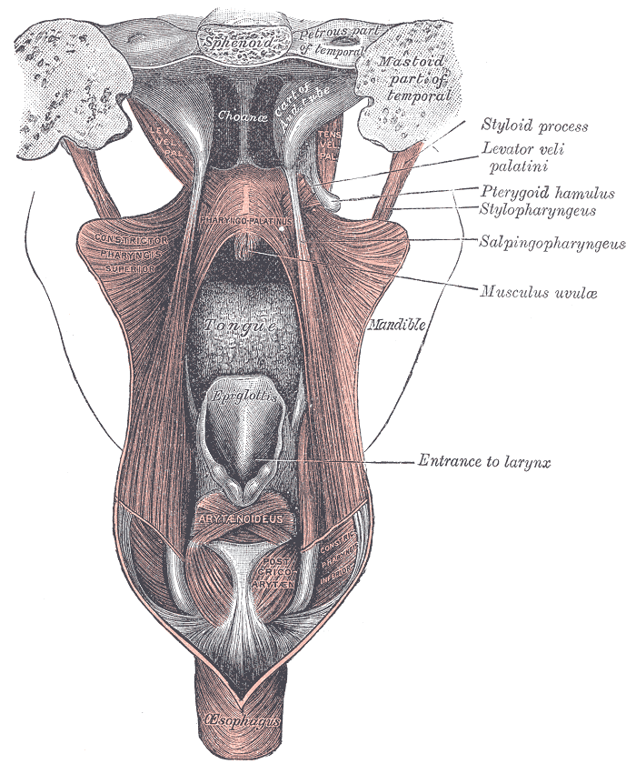
Soft Palate & Fauces (Mouth & Throat)
Palatine aponeurosis: * Levator veli palatini: Elevates the soft palate during swallowing.
Tensor veli palatini: Tenses the soft palate.
Musculus uvulae: Elevates and shortens the uvula.
Palatopharyngeus: Tenses the soft palate and pulls the pharynx upward.
Neck
General
Platysma: Tenses the skin of the neck and depresses the lower jaw.
Longus colli: Flexes and rotates the neck.
Longus capitis: Flexes the head at the neck.
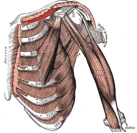
Scalenus anterior: Flexes the neck laterally and elevates the 1st rib.
Scalenus medius: Flexes the neck laterally and elevates the 1st rib.
Scalenus posterior: Flexes the neck laterally and elevates the 2nd rib.
(Scalenus minimus): Elevates the pleural dome.
Sternocleidomastoid: Rotates the head to the opposite side and flexes the neck.
Sub-occiput
Rectus capitis anterior: Flexes the head at the atlanto-occipital joint.
Rectus capitis lateralis: Flexes the head laterally.
Rectus capitis posterior major: Extends and rotates the head.
Rectus capitis posterior minor: Extends the head.
Obliquus capitis superior: Extends the head and flexes it laterally.
Obliquus capitis inferior: Rotates the head.
Supra-hyoid
Digastric: Depresses the mandible and elevates the hyoid bone.
Stylohyoid: Elevates and retracts the hyoid bone.
Mylohyoid: Elevates the hyoid and the floor of the mouth.
Geniohyoid: Pulls the hyoid bone forward and depresses the mandible.
Infra-hyoid
Sternohyoid: Depresses the hyoid bone.
Omohyoid: Depresses and retracts the hyoid bone.
Sternothyroid: Depresses the larynx (thyroid cartilage).
Thyrohyoid: Depresses the hyoid bone and elevates the larynx.
(Levator glandulae thyroideae): #### Cervical
Investing layer / Pretracheal layer / Prevertebral layer: * Carotid sheath: #### Pharyngeal
Pharyngeal raphe / Pterygomandibular raphe: * Superior/Middle/Inferior constrictor: Constricts the pharyngeal wall during swallowing.
Stylopharyngeus: Elevates and widens the pharynx.
Salpingopharyngeus: Elevates the pharynx.
Peripharyngeal space / Buccopharyngeal fascia: #### Laryngeal
Cricothyroid: Tenses and elongates the vocal cords (raises pitch).
Posterior crico-arytenoid: Abducts the vocal cords (opens the airway).
(Ceratocricoid): * Lateral crico-arytenoid: Adducts the vocal cords (closes the airway).
Vocalis: Adjusts the tension of the vocal folds for speech.
Thyro-arytenoid: Relaxes the vocal folds.
Oblique / Transverse arytenoid: Adducts the arytenoid cartilages to close the glottis.
-
Upper Back
Trapezius: Elevates, rotates, and retracts the scapula.
(Transversus nuchae): * Latissimus dorsi: Extends, adducts, and medially rotates the arm.
Rhomboid major / minor: Retracts and stabilizes the scapula.
Levator scapulae: Elevates the scapula.
Serratus posterior inferior: Depresses the lower ribs.
Serratus posterior superior: Elevates the upper ribs.
Anterior / Lateral posterior cervical intertransversarii: Assists in lateral flexion of the neck.
Intertransversarii laterales lumborum: Assists in lateral flexion of the spine.
Nuchal fascia: ### Lower Back
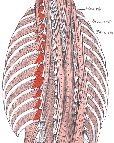
Erector Spinae: Primary extensor of the vertebral column.
Spino-transversales: Extends and rotates the head and neck.
Transverso-spinales: Extends and rotates the vertebral column.
Inter-spinales: Extends the vertebral column.
Inter-transversarii: Lateral flexion of the spine.
Thoracolumbar fascia: ---
-
Thorax
(Sternalis): * Pectoralis major: Adducts, flexes, and medially rotates the arm.
Pectoralis minor: Protracts and depresses the scapula.
Subclavius: Depresses and stabilizes the clavicle.
Serratus anterior: Protracts the scapula and stabilizes it against the ribs.
Levatores costarum: Elevates the ribs.
External intercostal muscle: Elevates the ribs during inhalation.
Internal intercostal muscle: Depresses the ribs during forced exhalation.
Innermost intercostal muscle: Assists the internal intercostals.
Subcostales: Depresses the ribs.
Transversus thoracis: Depresses the costal cartilages.
Pectoral / Clavipectoral / Thoracic / Endothoracic fascia: * Diaphragm: The primary muscle for inspiration (expands the chest cavity).
Abdomen
Rectus abdominis: Flexes the trunk (vertebral column).
Pyramidalis: Tenses the linea alba.
External oblique: Flexes and rotates the trunk.
Superficial inguinal ring: * Internal oblique: Flexes and rotates the trunk.
Transversus abdominis: Compresses the abdominal contents (core stability).
Linea alba / Linea semilunaris: Inguinal canal: Quadratus lumborum: Flexes the spine laterally and stabilizes the pelvis.
Abdominal / Pelvic fascia: * Pelvic diaphragm: Supports the pelvic viscera.
Perineal muscles: Supports the pelvic floor and helps control sphincters.
-
Upper Limbs
Deltoid: Primary abductor of the arm; flexes and extends the arm.
Supraspinatus: Initiates arm abduction and stabilizes the shoulder.
Infraspinatus: Externally rotates the arm.
Teres minor: Externally rotates the arm.
Teres major: Adducts and medially rotates the arm.
Subscapularis: Medially rotates the arm.
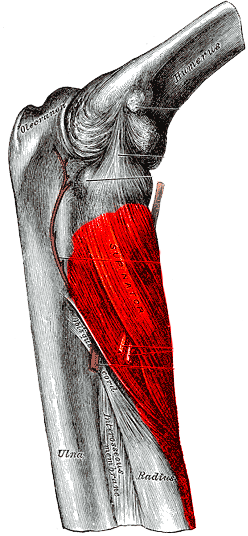
Biceps brachii: Flexes the elbow and supinates the forearm.
Coracobrachialis: Flexes and adducts the arm at the shoulder.
Brachialis: Primary flexor of the elbow.
Triceps brachii: Primary extensor of the elbow.
Anconeus: Assists in elbow extension.
Articularis cubiti: Pulls the joint capsule away during extension.
Pronator teres: Pronates the forearm.
Flexor carpi radialis: Flexes and abducts the wrist.
Palmaris longus: Flexes the wrist (weakly).
Flexor carpi ulnaris: Flexes and adducts the wrist.
Flexor digitorum profundus: Flexes the fingers and wrist.
Flexor pollicis longus: Flexes the thumb.
Pronator quadratus: Pronates the forearm.
Brachioradialis: Flexes the elbow.
Extensor carpi radialis longus / brevis: Extends and abducts the wrist.
Extensor digitorum: Extends the fingers and wrist.
Extensor digiti minimi: Extends the little finger.
Extensor carpi ulnaris: Extends and adducts the wrist.
Supinator: Supinates the forearm.
Abductor pollicis longus: Abducts and extends the thumb.
Extensor pollicis brevis / longus: Extends the thumb.
Extensor indicis: Extends the index finger.
Palmaris brevis: Tenses the skin of the palm.
Abductor pollicis brevis: Abducts the thumb.
Flexor pollicis brevis: Flexes the thumb.
Opponens pollicis: Opposes the thumb (moves it toward other fingers).
Adductor pollicis: Adducts the thumb.
Abductor digiti minimi: Abducts the little finger.
Flexor digiti minimi brevis: Flexes the little finger.
Opponens digiti minimi: Opposes the little finger.
Lumbricales: Flexes the knuckles and extends the fingers.
Dorsal interossei: Abducts the fingers (spreads them).
Palmar interossei: Adducts the fingers (pulls them together).
Lower Limbs
Iliopsoas: Primary flexor of the hip.
(Psoas minor): Assists in flexing the pelvis.
Gluteus maximus: Primary extensor and external rotator of the hip.
Gluteus medius: Abducts the hip and stabilizes the pelvis.
Gluteus minimus: Abducts and medially rotates the hip.
Gluteal aponeurosis: * Tensor fascia latae: Abducts and flexes the hip.
Piriformis: Externally rotates the abducted hip.
Obturator internus: Externally rotates the hip.
Gemellus superior / inferior: Externally rotate the hip.
Quadratus femoris: Externally rotates the hip.
Sartorius: Flexes, abducts, and externally rotates the hip; flexes the knee.
Quadriceps femoris: Primary extensor of the knee.
Articularis genus: Pulls the knee joint capsule upward during extension.
Pectineus: Adducts and flexes the hip.
Adductor longus / brevis / magnus / minimus: Adducts the thigh.
Gracilis: Adducts the thigh and flexes the knee.
Obturator externus: Externally rotates the hip.
Biceps femoris: Flexes the knee and extends the hip.
Semitendinosus: Flexes the knee and extends the hip.
Semimembranosus: Flexes the knee and extends the hip.
Tibialis anterior: Dorsiflexes and inverts the foot.
Extensor digitorum longus: Extends the toes and dorsiflexes the foot.
Fibularis tertius: Dorsiflexes and everts the foot.
Extensor hallucis longus: Extends the big toe and dorsiflexes the foot.
Fibularis longus / brevis: Plantarflexes and everts the foot.
Triceps surae (Calves): Primary plantarflexor of the foot (standing on toes).
Plantaris: Assists in plantarflexion and knee flexion.
Popliteus: Unlocks the knee joint to initiate flexion.
Tibialis posterior: Plantarflexes and inverts the foot.
Flexor digitorum longus: Flexes the toes.
Flexor hallucis longus: Flexes the big toe.
Extensor hallucis brevis: Extends the big toe.
Extensor digitorum brevis: Extends the toes.
Abductor hallucis: Abducts the big toe.
Flexor hallucis brevis: Flexes the big toe.
Adductor hallucis: Adducts the big toe.
Abductor digiti minimi: Abducts the little toe.
(Opponens digiti minimi): Opposes the little toe.
Flexor digiti minimi brevis: Flexes the little toe.
Flexor digitorum brevis: Flexes the toes.
Quadratus plantae: Assists in flexing the toes.
Lumbricals: Flexes the toes at the base and extends the tips.
Dorsal interossei: Abducts the toes.
Plantar interossei: Adducts the toes.
References:
Category:human muscles by body part (no date) Wikimedia Commons. Available at: https://commons.wikimedia.org/wiki/Category:Human_muscles_by_body_part (Accessed: March 23, 2023).
FCAT (Federative Committee on Anatomical Terminology), (1998) Terminologia Anatomica: International Anatomical Terminology, Stuttgart, Thieme Publishing Group.